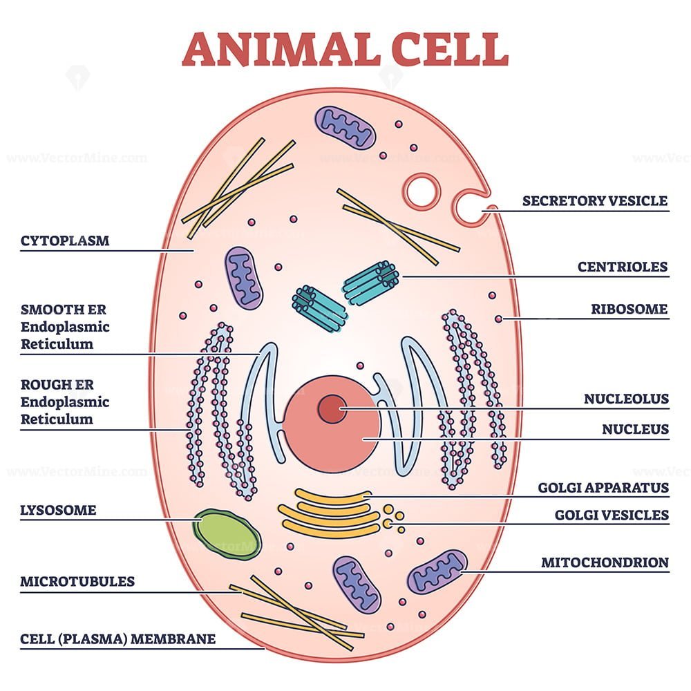
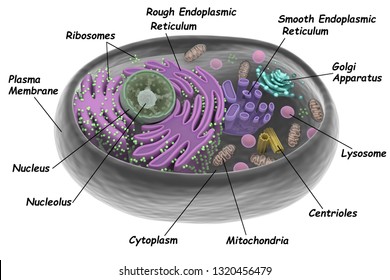
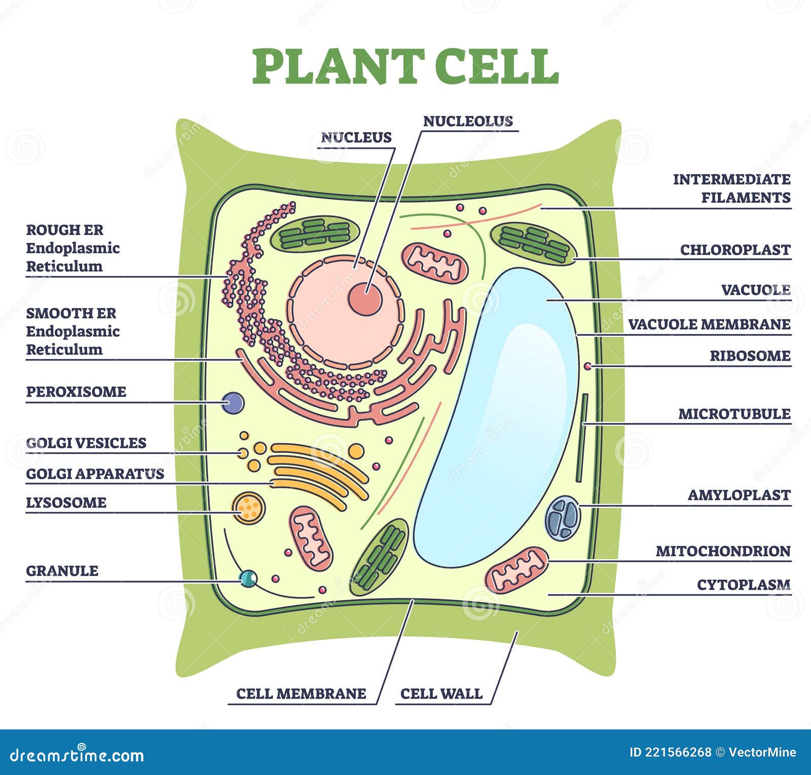
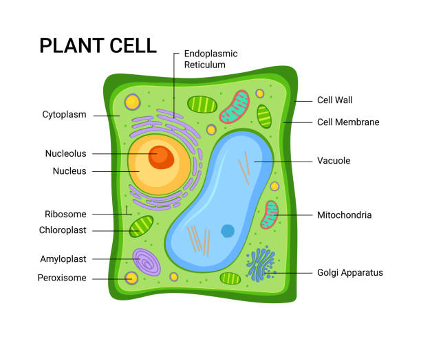
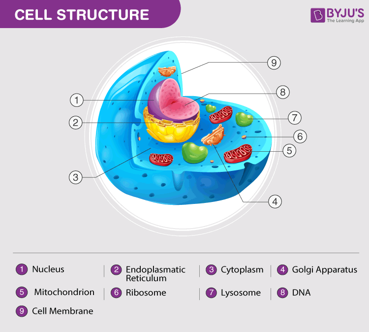
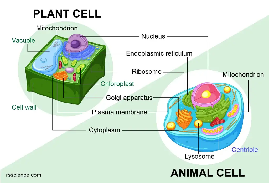
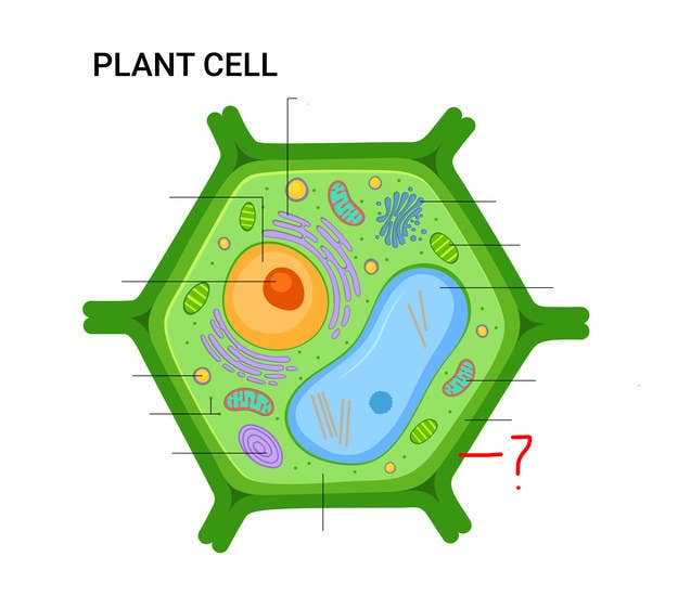
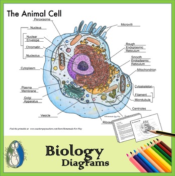
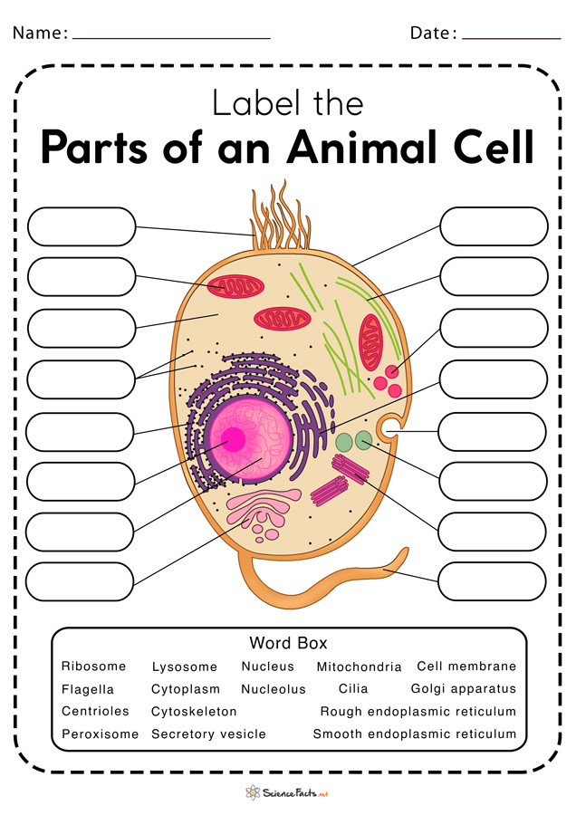
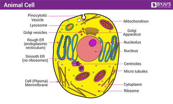
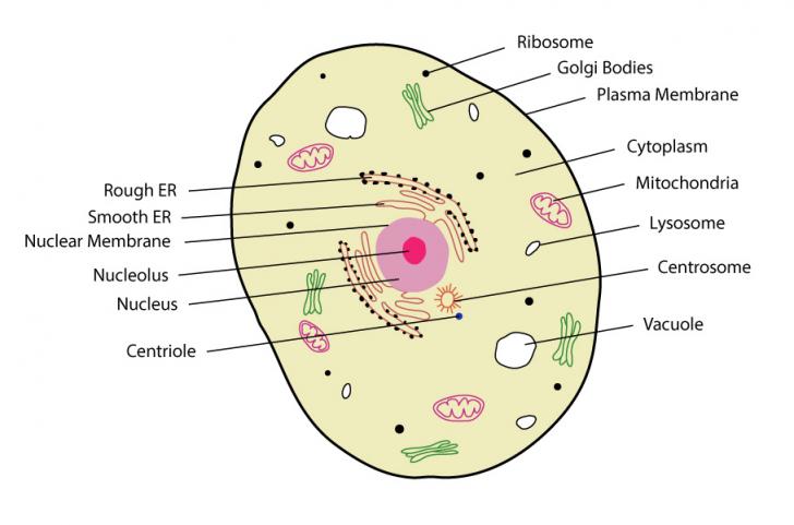
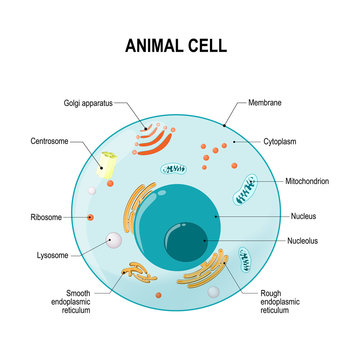
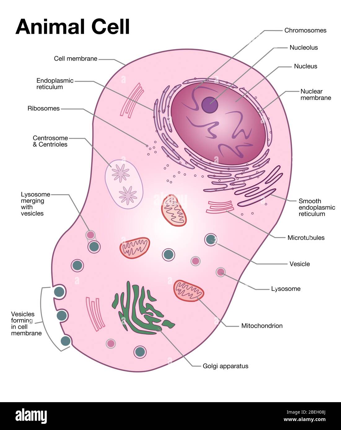
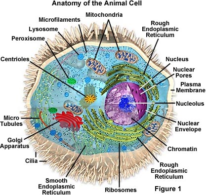
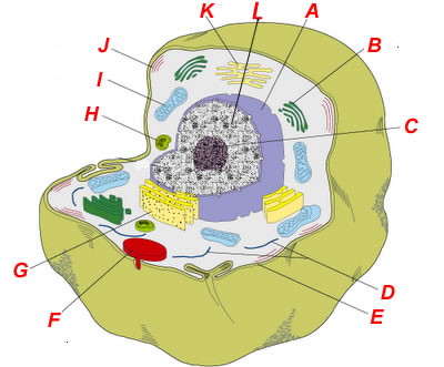
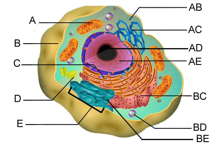
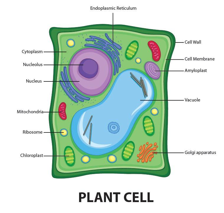
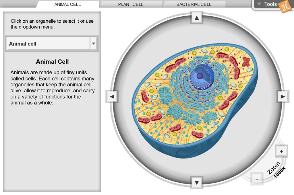
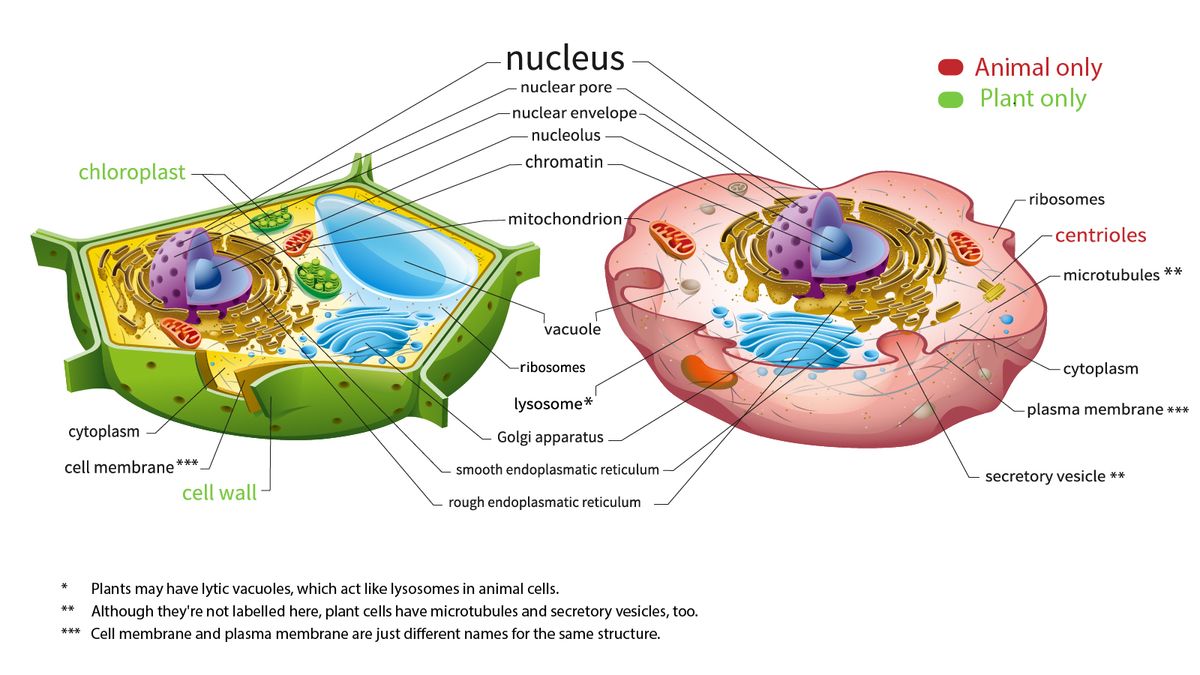
38 cell diagram and labels
Essential JS 2 for Angular - Syncfusion Essential JS 2 for Angular is a modern JavaScript UI toolkit that has been built from the ground up to be lightweight, responsive, modular and touch friendly. It is written in TypeScript and has no external dependencies. Action potential - Definition, Steps, Phases | Kenhub Action potential curve and phases (diagram) Hypopolarization is the initial increase of the membrane potential to the value of the threshold potential. The threshold potential opens voltage-gated sodium channels and causes a large influx of sodium ions. This phase is called the depolarization.
NCERT Book Class 8 Science Chapter 8 Cell - Structure and Functions January 7, 2022. in 8th Class. Reading Time: 3 mins read. 0. NCERT Book for Class 8 Science Chapter 8 Cell - Structure and Functions is available for reading or download on this page. Students who are in Class 8 or preparing for any exam which is based on Class 8 Science can refer NCERT Science Book for their preparation.

Cell diagram and labels
Self-Taught AI May Have a Lot in Common With the Human Brain Such "supervised" training requires data laboriously labeled by humans, and the neural networks often take shortcuts, learning to associate the labels with minimal and sometimes superficial... Class Diagram Relationships in UML Explained with Examples - Creately Blog Class diagrams are the main building block in object-oriented modeling. They are used to show the different objects in a system, their attributes, their operations, and the relationships among them. The following figure is an example of a simple class: Simple class diagram with attributes and operations Dendrites: definition, structure and function | GetBodySmart Dendrites are often, but not always, branched cytoplasmic processes that attach to one side of the cell body. They contain many of organelles found in the cell body, which perform the same metabolic functions. Dendrites make up most of the receptive zone of a neuron and can detect specific changes in the surrounding environment (stimuli).
Cell diagram and labels. Symmetry in Crystallography Notes - University of Oklahoma Unit Cells. These regularly-repeating blocks are now known as unit cells. ... If the structure is determined to have the incorrect handedness, then the structure is simply inverted through some symmetry point such as (0, 0, 0) or (0.5, 0.5, 0.5) and the refinement is completed. The space group must also be changed if the structure crystallizes ... The Cell - ScienceQuiz.net Two of the labels are incorrect. What are they?? Vacuole and chloroplast? Vacuole and cytoplasm? Nucleus and chloroplast? Cell wall and cell membrane; The diagram shows a plant cell. Identify parts A and B.? A = cell wall and B = nucleus.? A = cell wall and B = cytoplasm.? A = cell membrane and B = nucleus.? A = vacuole and B = nucleus. Look at the diagram of an … › cells › bactcellInteractive Bacteria Cell Model - CELLS alive In the space are enzymes and other proteins that help digest and move nutrients into the cell. Cell Wall: Composed of peptidoglycan (polysaccharides + protein), the cell wall maintains the overall shape of a bacterial cell. The three primary shapes in bacteria are coccus (spherical), bacillus (rod-shaped) and spirillum (spiral). Add vertical line to Excel chart: scatter plot, bar and line graph Select your source data and make a line graph ( Inset tab > Chats group > Line ). Set up the data for the vertical line in this way: In one cell (E1), type the text label for the data point at which you want to draw a line exactly as it appears in your source data.
Cell Membrane (Plasma Membrane) - Genome.gov The cell membrane, also called the plasma membrane, is found in all cells and separates the interior of the cell from the outside environment. The cell membrane consists of a lipid bilayer that is semipermeable. The cell membrane regulates the transport of materials entering and exiting the cell. Narration 00:00 … chemostratigraphy.com › how-to-plot-a-ternaryHow to plot a ternary diagram in Excel - Chemostratigraphy.com Sep 14, 2022 · Adding labels to the apices. Next, we need some space for the apices labels: click into the Plot Area (not the Chart Area) then resize by holding the Shift key (this ensures an equal scaling) and use the mouse cursor on one of the corner pick-points. Then recentre the Plot Area in the Chart Area. sciencequiz.net › newjcscience › jcbiologyThe Cell - ScienceQuiz.net A is the cell wall and DNA is located inside B. A is the cytoplasm and animal cells may have small vacuoles. A is the cell membrane and B contains chlorophyll. Feynman diagram - Wikipedia In theoretical physics, a Feynman diagram is a pictorial representation of the mathematical expressions describing the behavior and interaction of subatomic particles.The scheme is named after American physicist Richard Feynman, who introduced the diagrams in 1948.The interaction of subatomic particles can be complex and difficult to understand; Feynman diagrams give a simple visualization of ...
› cell_cycle_jsInteractive Cell Cycle - CELLS alive INTERPHASE. Gap 0. Gap 1. S Phase. Gap 2. MITOSIS . ^ Cell Cycle Overview Cell Cycle Mitosis > Meiosis > Get the Cell Division PowerPoints Animal Cell Diagram For Class 9 With Label Diagram To draw a well labelled diagram of an animal cell, the cell membrane has to be drawn. the cell membrane is an integral part of the cell structure that keeps the entire cell bound together. it is a phospholipid bilayer surrounding the entire cell with cytoplasm and the organelles in the cell cytoplasm. the membrane is selectively permeable only. IEC 60617 - Graphical Symbols for Diagrams IEC 60617 contains graphical symbols for use in electrotechnical diagrams. All the parts (Ed. 2 or 3) of the previously published IEC 60617 have been incorporated into this database that currently includes some 1900 symbols. The database is the official source of IEC 60617. Each graphical symbol is identified by a reference number and contains ... byjus.com › biology › skin-diagramSkin Diagram with Detailed Illustrations and Clear Labels - BYJUS Skin Diagram The largest organ in the human body is the skin, covering a total area of about 1.8 square meters. The skin is tasked with protecting our body from external elements as well as microbes.
20 Main Motherboard Components and Their Functions - MiniTool According to Wikipedia, a motherboard is the main printed circuit board (PCB) in common computers (both desktops and laptops) as well as other expandable systems. It is also known as a mainboard, main circuit board, system board, baseboard, logic board, planar board or mobo. Motherboard Components and Their Functions
'Mystery gene' matures the skeleton of the cell -- ScienceDaily Cell biologists are very interested in actin, because actin -- a protein of which we produce more than 100 kilograms in our lifetime -- is a main component of the cell skeleton and one of the most ...
An introduction to the SingleCellExperiment class - Bioconductor A common procedure in single-cell analyses is to identify relationships between pairs of cells, e.g., to construct a nearest-neighbor graph or to mark putative physical interactions between cells. We can capture this information in the SingleCellExperiment class with the colPairs () functionality.
Circulatory system - Wikipedia Diagram of the human heart showing blood oxygenation to the pulmonary and systemic circulation The heart pumps blood to all parts of the body providing nutrients and oxygen to every cell, and removing waste products. The left heart pumps oxygenated blood returned from the lungs to the rest of the body in the systemic circulation.
Fungal cells: characteristics, organelles and functions Fungal cells are characterized by being elongated and tubular in shape, with rounded edges. They have a cell wall. Like plant cells, fungal cells are surrounded by a rigid structure known as the cell wall, which helps protect the cell, give it support and a defined shape. This cell wall is made up of a carbohydrate called chitin. Make up hyphae
Making Your Own Plant Cell Model At Home Is THIS Easy Cell wall - A cardboard box or foam board to make sides and base Green paint Cell membrane - Yellow card or white card and yellow paint Cytoplasm - Black paint or black foam board. You could use bubble wrap, painted black. Nucleus - Tennis ball Blue paint Hot glue gun and glue
WHMIS 2015 - Pictograms : OSH Answers - Canadian Centre for ... The product label and Section 2 (Hazards Identification) of the SDS still require the signal word, hazard statement (s), and other required label elements. WHMIS 2015 classes and categories that do not require a pictogram are: Flammable gases - Category 2 Flammable liquids - Category 4 Self-reactive substances and mixtures - Type G
byjus.com › biology › diagram-of-neuronA Labelled Diagram Of Neuron with Detailed Explanations - BYJUS Diagram Of Neuron with Labels. Here is the description of human neuron along with the diagram of the neuron and their parts. The neuron is a specialized and individual cell, which is also known as the nerve cell. A group of neurons forms a nerve.
Interactive Bacteria Cell Model - CELLS alive Cell Wall: Composed of peptidoglycan (polysaccharides + protein), the cell wall maintains the overall shape of a bacterial cell. The three primary shapes in bacteria are coccus (spherical), bacillus (rod-shaped) and spirillum (spiral). Mycoplasma are bacteria that have no cell wall and therefore have no definite shape. Outer Membrane: This lipid bilayer is found in Gram negative …
Diagram of Human Heart and Blood Circulation in It A heart diagram labeled will provide plenty of information about the structure of your heart, including the wall of your heart. The wall of the heart has three different layers, such as the Myocardium, the Epicardium, and the Endocardium. Here's more about these three layers. Epicardium
Science A-Z Science Diagrams - Visual Teaching Tools Science Diagrams from Science A-Z provide colorful, full-page models of important, sometimes complex science concepts. Science Diagrams, available in both printable and projectable formats, serve as instructional tools that help students read and interpret visual devices, an important skill in STEM fields.
Bronchioles: function and diagram | GetBodySmart An enlarged cross-sectional view of a bronchiole reveals the tissue layers that make up the wall. 1. 2. Ciliated simple columnar cells form the epithelial lining in the large bronchioles. In the small bronchioles, the epithelium changes to simple cuboid cells. Goblet cells and seromucous glands become less numerous with each bronchiole division.
Simple Staining: Principle, Procedure, Uses - Microbe Online The simple stain can be used as a quick and easy way to determine the cell shape, size, and arrangements of bacteria. True to its name, the simple stain is a very simple staining procedure involving a single solution of stain. Any basic dye such as methylene blue, safranin, or crystal violet can be used to color the bacterial cells.
Make Pareto chart in Excel - Ablebits.com By default, a Pareto graph in Excel is created with no data labels. If you'd like to display the bar values, click the Chart Elements button on the right side of the chart, select the Data Labels check box, and choose where you want to place the labels: The primary vertical axis showing the same values has become superfluous, and you can hide it.
Interactive Cell Cycle - CELLS alive INTERPHASE. Gap 0. Gap 1. S Phase. Gap 2. MITOSIS . ^ Cell Cycle Overview Cell Cycle Mitosis > Meiosis > Get the Cell Division PowerPoints
ej2.syncfusion.com › angular › demosEssential JS 2 for Angular - Syncfusion Essential JS 2 for Angular is a modern JavaScript UI toolkit that has been built from the ground up to be lightweight, responsive, modular and touch friendly. It is written in TypeScript and has no external dependencies.
Mr. Jones's Science Class Cells Cells Overview Sheet Cells - Self-Assessment Sheet Prokaryotes vs. Eukaryotes Celley's Trip to Cell City Cell City Map (PPT.) Cell Parts Activity Cells - Outline Cell Diagrams Cell Parts - Analogies Cells - Identifying Words with Pictures Cell Structure and Function Crossword Photosynthesis vs. Cellular Respiration Cell-O Cell Art Find ...
Single-crystal X-ray Diffraction - Techniques The primitive unit cell is refined using least-squares and then converted to the appropriate crystal system and Bravias lattice. This new cell is also refined using least-squares to determine the final orientation matrix for the sample. After the refined cell and orientation matrix have been determined, intensity data is collected.
Plasma Membrane (Cell Membrane) - Genome.gov The plasma membrane, also called the cell membrane, is the membrane found in all cells that separates the interior of the cell from the outside environment. In bacterial and plant cells, a cell wall is attached to the plasma membrane on its outside surface. The plasma membrane consists of a lipid bilayer that is semipermeable.
Spinal Cord Diagram with Detailed Illustrations and Clear Labels Spinal Cord Diagram. The spinal cord is one of the most important structures in the human body. It is the most important structure for any vertebrate. Anatomically, the spinal cord is made up of nervous tissue and is integrated into the spinal column of the backbone. Main Article: Spinal Cord – Anatomy, Structure, Function, and Spinal Cord Nerves; Also Read: Central Nervous System ...
Skin Diagram with Detailed Illustrations and Clear Labels - BYJUS Skin Diagram. The largest organ in the human body is the skin, covering a total area of about 1.8 square meters. The skin is tasked with protecting our body from external elements as well as microbes. Interesting Note: The skin is also responsible for maintaining our body temperature – this was apparent in victims who were subjected to the medieval torture of being skinned alive. …
How to Test Graphs and Charts (Sample Test Cases) - Software Testing Help Sample Test Cases for Testing Graphs and Charts. 1) No data found message should be displayed when there is no data in the graph. 2) Waiting cursor or Progress bar should be given on Graph Load. 3) Correct values displayed with respect to its Pivot table (values of the graph x-axis & y-axis matches its table values) 4) If the Graph is ...
2D Semantic Labeling - International Society for Photogrammetry and ... Focus is on detailed 2D semantic segmentation that assigns labels to multiple object categories. Further research drivers are very high-resolution data from new sensors and advanced processing techniques that rely on increasingly mature machine learning techniques. Despite the enormous efforts spent, these tasks cannot be considered solved, yet.
How to plot a ternary diagram in Excel - Chemostratigraphy.com 14/09/2022 · Ternary diagrams are common in chemistry and geosciences to display the relationship of three variables.Here is an easy step-by-step guide on how to plot a ternary diagram in Excel. Although ternary diagrams or charts are not standard in Microsoft® Excel, there are, however, templates and Excel add-ons available to download from the internet.
Excel Waterfall Chart: How to Create One That Doesn't Suck - Zebra BI The legend, the vertical axis and labels, the horizontal grid lines - none of them contribute to the reader's better understanding of the data. If anything, they are a distraction. So, let's remove all unnecessary elements and write our key message to the title. It's a shame that the chart title cannot be inserted automatically from a cell.
Circulatory System Diagram | New Health Advisor Coronary circuit mainly consists of cardiac veins including anterior cardiac vein, small vein, middle vein and great (large) cardiac vein. There are different types of circulatory system diagrams; some have labels while others don't. The color blue stands for deoxygenated blood while red stands for blood which is oxygenated.
Dendrites: definition, structure and function | GetBodySmart Dendrites are often, but not always, branched cytoplasmic processes that attach to one side of the cell body. They contain many of organelles found in the cell body, which perform the same metabolic functions. Dendrites make up most of the receptive zone of a neuron and can detect specific changes in the surrounding environment (stimuli).
Class Diagram Relationships in UML Explained with Examples - Creately Blog Class diagrams are the main building block in object-oriented modeling. They are used to show the different objects in a system, their attributes, their operations, and the relationships among them. The following figure is an example of a simple class: Simple class diagram with attributes and operations
Self-Taught AI May Have a Lot in Common With the Human Brain Such "supervised" training requires data laboriously labeled by humans, and the neural networks often take shortcuts, learning to associate the labels with minimal and sometimes superficial...














Post a Comment for "38 cell diagram and labels"